Thursday, August 29, 2019

MATLAB exercises - #1

First part of MATLAB practice exercises

  1. Generate random data for testing purpose
  2. Output mean and standard deviation of the data
  3. Plot histogram of the data

Part 1.

Generate random data:

  • using uniform distribution rand()
  • using normal distribution randn()

Using uniform distribution:

x_uniform = rand(100,1); % creates vector of 100 elements samples from uniform distribution, 0<=x<=1

Using uniform distribution:

x_normal = randn(100,1); % creates vector of 100 elements samples from normal distribution

Part 2.

Output mean of x_uniform:

mean(x_uniform)

Now, output mean of x_normal:

Show answer
mean(x_normal)

Output standard deviation of x_uniform:

std(x_uniform)

Now, output standard deviation of x_normal:

Show answer
mean(x_normal)

Part 3.

Plot histogram of the data using hist()function. Do it for both data vectors (random data from uniform and normal distributions)

Show answer
x_uniform = randn(100,1);
hist(x_uniform)


Sunday, August 25, 2019

The Cell 9-8

The Cell (Alberts) 9-8 question Antibodies that bind to specific proteins are important tools for defining the locations of molecules in cells. The sensitivity of primary antibody, the antibody that reacts with the target molecule, is often enhanced by using labeled secondary antibodies that bind to it. What are the advantages and disadvantages of using secondary antibodies that carry fluorescent tags versus those that carry bound enzymes? Show answer
A lot of useful information can be found on Wikipedia article Immunohistochemistry . Fluorescent tags on secondary antibody allow for higher signal and labeling multiple targets using orthogonal pairs of antibodies. For example, we can label at the same time tubulin and actin. Secondary antibodies, though, are bulkier and can decrease resolution, especially in super-resolution microscopy. Using multiple antibodies also can increase chance of off-target labeling (when antibody binds elsewhere rather than it's antigen).

Thursday, February 8, 2018

GRE Molecular & Cell BIology Practice test Q 76

Show answer

This question is concerned with organisms that can or cannot live without exogenous (added to the environment) chemical X. By definition auxotrophs are those that need some essential nutrient to be added, prototrophs can synthesize their own chemical X. Also, we need to know what is

"complementation group". By definition, that is pair of mutants that have same phenotype (say, they are auxotrophs for nutrient X), but when crossed will produce wild-type (prototroph) progeny. That means that mutations in these mutants occurred in different genes, and each parent "complemented" each other, supplied wild-type gene to correct for another parent's mutation. "Complementation group" is a group of genes that do not compliment each other. By definition, each gene can be in its own complementation group.

From data, mutants 2 and 4 do not complement each other, so are mutants 3 and 5. All what's left is mutant 1, which by definition doesn't complement itself, hence in its own group.

 Answer is C

 This is also a useful link: http://courses.washington.edu/gensc371/lecture/jan22.pdf

Sunday, February 4, 2018

GRE Molecular & Cell Biology Questions 170, 171, 172


Today question is from GRE Biochemistry, Cell and Molecular Biology practice test.
Show answer

A1. From Fig. 1&2 we can see that protein synthesis continues for some time after inhibitor was added. That is caused by the fact that even when inhibitor is added and significant fraction of RNA Polymerase activity is halted, there is still a lot of "idle" or not yet used products of RNA Pol in the cell. This backup amount of RNAs is available for protein synthesis for short time. Answer is A.

A2. From Fig. 1 we can approximate rate of synthesis after inhibitor was added, and compare it to non-inhibited case. TO do that, let's just calculate derivative after inhibition, or slope of the lines. Take vertical distance (change in Counts Per minute, CPM), divide by change in time.

No inhibition: 6000-2000 / 20 hrs ~ 4000CPM/20min=200CPM/hr
With inhibition: 3000-2000 / 20 hrs ~ 50CPM/hr

After inhibition, rate goes down about 4x, thus we can estimate that inhibitor blocks about 75% of activity. Answer is B.

A3. In this question experiment was performed as following: proteins from nuclear extract were purified on a column. Peak were collected and used as a source for RNA polymerization. Since radioactive thymine was used, RNA polymerization activity of each fraction (peak) was measured afterwards.

In HeLa cells, there are 3 RNA polymerases:
RNA Pol I: synthesizes a pre-rRNA 45S, molecular weight 590 kDa
RNAPol II: synthesizes precursors of mRNAs and most snRNA and microRNAs, MW 550 kDa
RNA Pol III: synthesizes tRNAs, rRNA 5S and other small RNAs found in the nucleus and cytosol, MW 0.7MDa

Question is about type of the RNA Pol that is concentrated in first peak. Interestingly, Fig 3 gives much more information that asked in question, possibly setting up test-takers for a distraction. To answer, I had to Google RNA Pol chromatography, and it seems that peak 1 is mainly RNA Pol I.
 

 But even more widely known is that nucleolus is a site where ribosomal RNA (rRNA) is primarily produced. Answer is A.

Sunday, March 19, 2017

Bonus day: question 36 from Physics GRE

This question comes from practice Physics GRE exam (2011 edition)

The capacitor in the circuit above is charged. If switch S is closed at time t = 0, which of the following represents the magnetic energy, U, in the inductor as a function of time? (Assume that the capacitor and inductor are ideal.)

That's a relatively simple question with P+=50%. Now let's get to the answer: Show answer
Correct answer is A

To get to it we might use process of elimination: find all answers that are NOT correct and see what's left. First of all, question says "Assume that the capacitor and inductor are ideal". What that means right off the bat is that there should be no dissipation of energy. So answer D is incorrect right away. Also, we should remember that inductor's energy depends on current (contrast it to the fact that capacitor's energy depends on voltage across its plates). In initial conditions there is no current going thorugh the circuit because switch S is open. Hence, correct answer should have plot start at 0. This eliminates answers B, C, D (again). We are left with either E or A answers. You can pick one by again referring to ideal-ness or ideality of the circuit. Indefinite oscillations (answer A) are the only possibility in that case. By the way, the fact that this quite simple question has P+ of just 50% should tell you that it is not that hard to score high on Physics GRE. You can do it!

Sample GRE Q55

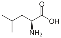
Turns out this blog is still up and running :-) so here is a new one: Sample GRE biochem test, question 55 with P+ of 31% (from 2016 edition) [*P+ is percentage of test takers who gave correct answer.]

An amino acid transporter protein is responsible for the transport of a specific amino acid across a membrane. The KI values of several competitive inhibitors of the amino acid transporter are shown above. Based on these data, which of the following is most likely the amino acid transported by this protein?


Show answer
Correct answer is E And here is why. First, let's unwrap some of the language. "The inhibitor constant, Ki, is an indication of how potent an inhibitor is; it is the concentration required to produce half maximum inhibition." (here) So, the higher the Ki, more inhibitor concentration is necessary to prevent our transporter from moving amino-acid X through the membrane. It is also mentioned in the question that inhibitors are competitive. That means, to some degree, that inhibitors are similar in structure to that of the amino-acid X. Most similar inhibitor will then have lowest Ki, is est will occupy transporter's "slot" very nicely preventing amino-acid X from binding. In other words, inhibitor with lowest Ki will have most similar structure. Lowest Ki belongs to competitive inhibitor #3 (calledethyl benzene, by the way). It might be useful to notice second-best inhibitor as well, #5 (isoamyl alcohol). Both of these compounds are relatively not charged. Now let's look at structures of all possible answers:
(A)
(B)
(C)
(D)
(E)


As you can see, only tyrosine (E) carries benzyl group and also has less charge than other compounds. hence the answer is E

Saturday, December 21, 2013

Question #97 of GRE Sample Subject test (MolBio)

Propagation of a regenerative action potential
along an axon can be accelerated by which of the
following?
(A) A decrease in the transmembrane resistance
(B) A decrease in the axoplasmic resistance
(C) Reduced myelin wrapping
(D) Shortened internodal lengths
(E) Narrowing of the axon diameter

Show answer
Answer:
(B)

First of all, what "regenerative" action potential means? Kandel's textbook defines just an action potential (AP) as a regenerative electrical signal.
Now we can unwrap answers and say what will happen in each case.
(A) A decrease in the transmembrane resistance = essentially that is increase in channels number. But not Hodgkin's channels, but so-called "leaky" channels that prevent absolute ion separation by Na/K-ATPase. If leaky channels were more abundant (or more active, you can imagine some drug that does that) it would be more difficult for membrane to stay polarized.

(C) Reduced myelin wrapping = As we all know, myelin facilitates rapid AP propagation by essentially increasing membrane resistance. Thinning of myelin will slow down impulse propagation.

(D) Shortened internodal lengths = shortening of distance between ranvier nodes. This will also slow down signal propagation. There are two ways for AP to travel along axon: as it happens in umyelinated axons and "jumping" from one node to another (saltatory conduction). In latter case, because of myelin isolation (high resistance of membrane), depolarization at one node is enough to trigger channels' opening in nearby nodes (on distance ~200-2000  um). "Inside" nodes signal is propagated in general fashion: channels opens one by one in orderly manner and go through open-closed/inactivate-close/active cycle.
Shortening of internodal length will slow down signal. However, increase in distance between nodes might decrease sensitivity of axon to signal propagation. That is, not every single AP or not every packets of APs will propagate from one node to distal one, because depolarization will essentially be lost on the road due to diffusion of ions.

(E) Narrowing of the axon diameter = Seemingly useful change for rapid ion diffusion, that will not speed up signals. Axon diameter is very important for AP since it is resistance that prevent signal from travelling. Lower resistance can be achieved by increasing diameter or ions concentration. However, I suppose, that ion concentration change will affect channels activity and other physiological properties before change in conductivity. Ions here act like free electrons in semiconductors.

(B) A decrease in the axoplasmic resistance. Now, that is one correct answer. As discussed on (E) resistance of cytoplasm in axon (axoplasm, duh) plays important role in AP propagation.
Now, what is bad with answer (A)? Honestly, it was on my correct-answers-list for that question. if you think about it, axon consists of two parts: conductivity and depolarization mechanisms. Cytoplasm resistance contribute to conductivity, however leaky channels and voltage-dependent channels influence depolarization and AP generation. AP propagation does not really change with membrane resistance, but rather act as filter for action potentials that are too low. Or other wise, permit easily AP generation by keeping cell membrane closer to threshold (you know what that is, right?)

Monday, January 31, 2011

#15 RNA stem-loop stability

104. Which of the following mRNA molecules would form the most stable stem-loop structure?
(A) 5'...GGCUU......UUCGG...3'
(B) 5'...GGCUU......AAGCC...3'
(C) 5'...GGCUU......GGCUU...3'
(D) 5'...GGCUU......CCGAA...3'
(E) 5'...AAGCC......AAGCC...3'

Answer
First of all, stem-loops are formed by so-called inverted repeats. Is est, one strand of loop is complement to the other:
5'-...GGGCCC... (small loop here)
3'-...CCCGGG...
If this sequence is linearized now, we will get
5'-...GGGCCC......GGGCCC...-3' strand
Stem is that paired part of looped RNA.
Any mismatches (when, e.g. G is opposed by A) are called bulges.

So, hairpin (synonym of stem-loop) stability is defined by number of mismatches and stability of each base pair in the stem. That one arise from the fact that A have 2 hydrogen bonds with U (or T in DNA) and G have 3 hydrogen bonds with C, so GC pair is much more stable.

From first fact we can eliminate answer choices A, C, D and E, since neither A binds to C, nor U to U or A to U (just compare two nucleotides on the inner edges of uncovered sequences). Lets check answer B to be sure.
5'-...GGCUU...(loop)
3'-...CCGAA...
Should be stable!

This question is from easiest ones, so should not take a lot of time, which we are short of.

Yes, and I am back, will try to complete this blog during preparation.

Monday, December 14, 2009

#12 ER lumen protein

GRE-ST question number 54 is about ER proteins and functions.
The KDEL sequence, found on luminal proteins of the ER, is responsible for
(A) translocation of proteins into the ER lumen
(B) insertion of proteins into the membrane of the ER
(C) quality control in the ER
(D) recognition by signal peptidase of the signal sequence
(E) retrieval of ER luminal proteins from the Golgi

Show answer
This question is just seems to be hard. Just google and you'll probably find this wikipedia article on ER, where KDEL function is described clearly.
Point is that there are two types of proteins in ER lumen: those which should be transported further (to Goldgi) and those which work inside ER (e.g. linking oligosaccharides to proteins). So there should be some anchor for that "stable" proteins and it links to KDEL sequence on the end of those proteins. And because they cannot be fixed any way (or it would be very, very energetically unfavourable) they are transported back from Golgi to ER by specific KDEL-recetors.
So right answer is E.

Sunday, December 6, 2009

#14 Protein secretion

GRE-ST question #98.
All of the following processes occur in the pathway leading to regulated protein secretion in animal cells EXCEPT
(A) formation of transport vesicles from the rough endoplasmic reticulum
(B) an increase in the concentration of cytosolic calcium ions prior to secretion
(C) synthesis of an amino-terminal signal sequence
(D) phosphorylation of a mannose residue in a glycoprotein
(E) trimming of N-linked oligosaccharides
Show answer
That is pretty simple. Question can be solved by just following common protein secretion pathway. That starts by transcription DNA's gene into RNA. Then that RNA — called matrix RNA, mRNA — caught by ribosome — which is actually RNA too, but ribosomal thus rRNA — and with assistance of different translation factors protein production begins (that is called translation mRNA into protein). Why secretion proteins are selected ones? That is because they have special signal at the beginning of polypeptide chain on N-terminal (which is actually some sequence at the beginning of mRNA, of course) which is caught by special cytoplasmic protein — Signal Recognition Particle or SRP. When SRP bind to that signal sequence translation stops and ribosome with mRNA and bound SRP flows to endoplasmic reticulum, where SRP-receptors are located. They bind SRP and passes signal through the lipid bilayer of ER. Then translation continues and protein get into ER lumen. After that happened, protein undergo different saccharides modifications. Pathway of Asn modification clearly described in Alberts book, includes trimming of N-linked oligosaccharides. Main steps of protein movement are: secretion in vesicle from ER to Goldgi apparatus and same movement through different compartments of Goldgi apparatus. After that vesicle with protein inside fuse into cell's membrane thus secreting protein into extracellular space.
So, I suppose that no calcium used in this pathway except signal transduction to muscle cells. So the answer is B.